The California Institute for Regenerative Medicine awards $3.73 million to Mark Humayun, MD, PhD to develop a novel treatment for dry age-related macular degeneration

By Alexandra Demetriou

The California Institute for Regenerative Medicine (CIRM) recently awarded $3.73 million to Mark Humayun, MD, PhD, who serves as director of the USC Dr. Allen and Charlotte Ginsburg Institute for Biomedical Therapeutics and co-director of the USC Roski Eye Institute. The grant is part of CIRM’s translational research program to propel California forward as a hub of regenerative medicine breakthroughs. The funding will support researchers at the USC Ginsburg Institute as they develop a new treatment for dry age-related macular degeneration (dry AMD) –– a disease that has historically been considered difficult to treat.

This injection is not a man-made drug, but is instead composed of molecules produced by healthy retinal cells cultured in the laboratory. The researchers have already demonstrated that an intraocular injection of these molecules stimulates healing and slows down retinal degeneration in pre-clinical studies. The team’s prestigious CIRM grant is intended to accelerate the project’s transition to the clinical trial stage, thanks to its potential to benefit society by filling an unmet medical need.

One innovation inspires another

Approximately two million Americans currently live with advanced forms of AMD, and more than seven million deal with early-stage symptoms of the disease. Of those cases, 85-90% are the dry form of AMD. The condition is characterized by the progressive deterioration of the macula, or the region of the retina responsible for the clearest, most focused vision in the center of one’s field of view. A single layer of cells under the macula called the retinal pigment epithelium (RPE) is responsible for nurturing the eye’s photoreceptor cells, which lie directly on top of the RPE and perceive light.

As dry AMD progresses, patients perceive a dark spot obscuring the center of their vision. (Image: National Eye Institute, National Institutes of Health)

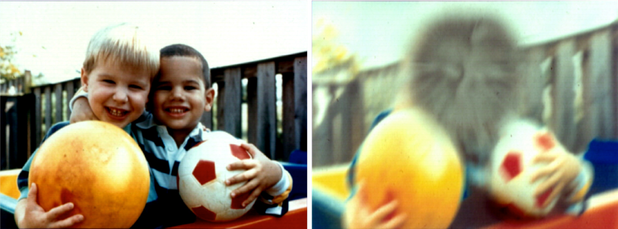
As the macula deteriorates and RPE cells die off, the photoreceptor cells necessary for vision gradually follow suit. Soon, patients begin to perceive a dark spot in the center of their visual field that can interfere with anything from reading to recognizing the faces of loved ones. The disease robs patients of both their eyesight and a great deal of their autonomy.

With the help of a CIRM Disease Team grant, a team of USC Ginsburg Institute researchers recently used stem cells to develop a retinal implant composed of a single layer of RPE cells to replace the degenerating part of the macula. Humayun, working alongside Amir Kashani, MD, PhD, surgically placed implants in 15 patients’ retinas. Although the trial was only intended to assess the implant’s safety, the team observed early signs of therapeutic benefit and a few patients even regained some of their eyesight. That project is currently poised to enter a larger clinical trial phase with the goal of becoming the first ever FDA-approved treatment for late-stage dry AMD.

While testing the implants in pre-clinical models, the team made an intriguing observation: the implanted cells had a restorative effect not only in the exact location they were placed, but also on the surrounding retinal tissue.

The researchers attributed this phenomenon to something they call the paracrine effect: cells from the implant produce chemical messages that communicate with surrounding cells –– a process known to biologists as “paracrine signaling” –– which causes the degenerating native cells to behave more like the healthy, newly implanted ones.

USC Ginsburg Institute researchers are culturing healthy retinal cells and harnessing the biological factors they produce to create therapeutic injections and reverse the symptoms of dry AMD. (Image: Kabir Ahluwalia)

The researchers asked themselves a key question: if these biological factors alone could have a restorative effect on a degenerating retina, could the team create an injectable solution containing the factors to complement the implant as an early-stage intervention for dry AMD?

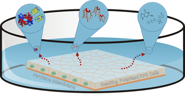
So far, the answer appears to be yes. Cells used in the implants are grown in the lab in a nutritious broth called the media, and as they grow, the molecular factors they produce are released into the media. By harvesting that factor-filled fluid and delivering it as an intraocular injection, the team has already seen success in dramatically slowing down the progression of retinal cell loss in pre-clinical models of retinal degeneration. The team’s long-term goal is to develop a therapeutic injection for early-stage patients to slow disease progression, while patients whose diseases have progressed to the point of blindness can receive implants.

Sights set on a future cure

Now that the researchers know this media holds promise as a therapeutic injection, the next step is to fully characterize its components. While the team has narrowed down the number of potentially therapeutic molecules in the media, no single constituent seems to have a strong effect on its own, meaning the factors may work together in a synergistic way to restore retinal function. Funding from the CIRM grant is meant to expedite the process of characterizing the media and creating a stable, reproducible therapeutic injection for eventual use in humans.

Kabir Ahluwalia, a doctoral student in the USC School of Pharmacy working on the project, explains that this research holds particular promise for those estimated seven million early-stage patients making up the majority of dry AMD cases. Early-stage AMD patients can currently benefit from specific nutritional supplements that slow down disease progression by about 25%, but many patients still need additional therapy. This novel injection of soluble growth factors could potentially serve as an ideal future treatment to prevent vision loss and provide renewed hope for these patients.

Dr. Mark Humayun featured in Nature Outlook for groundbreaking work to treat blindness

By Alexandra Demetriou

Dr. Mark Humayun from the USC Dr. Allen and Charlotte Ginsburg Institute for Biomedical Therapeutics was recently featured in Nature Outlook for his outstanding contributions to advance the treatment of blindness. The article highlighted a handful of the world’s top researchers tackling the problem of retinal degeneration, which is the leading cause of blindness in developed countries.

Image: Nature Outlook

Dr. Humayun’s Argus series implants were showcased amongst the most cutting-edge approaches to restoring eyesight for patients with some functional retinal cells still intact. The Argus II “bionic eye” consists of an electrode array that is implanted on the surface of the retina. The patient wears glasses equipped with a small video camera that transmits signals wirelessly to the implant. The electrodes stimulate the retina, which then communicates those signals to the brain. Over 300 patients have received the Argus II prosthesis and have regained their perception of light patterns, movement and basic shapes.

For patients who have completely lost functionality of the retina, Humayun and his colleagues at Second Sight have a different approach: sending signals from a camera directly to the brain.

The researchers have developed a chip, called Orion, which is surgically implanted on the outer surface of the very back of one’s brain. This region of the brain, called the visual cortex, is responsible for processing and interpreting information from the eyes. Like Argus II, Orion receives signals from a camera mounted on the patient’s glasses, and the brain can then convert those signals into visual information. So far, the chip has been successfully implanted in five patients with limited or no light perception. The trial is still its early stages, but the preliminary results look promising and Humayun hopes the chip will receive FDA approval in a few years.

To read the entire Nature Outlook article, click here.

Researchers at the USC Ginsburg Institute for Biomedical Therapeutics develop augmented reality glasses to help low-vision patients navigate their environments

By Alexandra Demetriou

A team of researchers at the USC Dr. Allen and Charlotte Ginsburg Institute for Biomedical Therapeutics recently developed a pair of augmented reality (AR) glasses to help visually impaired patients navigate their surroundings and perceive depth more clearly.

The glasses were designed to help patients with a degenerative eye disease called retinitis pigmentosa (RP). The condition causes progressive vision loss, particularly on the periphery of one’s vision, and makes it difficult to see in low-light conditions. Patients with RP often experience tunnel vision and have trouble perceiving their 3D environment. Specifically, they struggle to grasp objects and avoid obstacles in their path, and these issues are worse at night.

The AR glasses help solve this problem by allowing patients to see a color-coded mesh on top of the objects in their surroundings. The colors correspond to depth, with objects closest to the wearer appearing white, followed by green, blue and eventually red for objects that are furthest away.

table
The augmented reality glasses allow patients to perceive a color-coded mesh on top of the objects in their surroundings, which helps wearers perceive depth. (Image: Anastasios Nikolas Angelopoulos)

The device, customized by Anastasios Nikolas Angelopoulos and Dr. Mark Humayun, was configured with the user’s experience in mind. Rather than using virtual reality, which completely replaces the wearer’s field of view with an image on a screen, the augmented reality color mesh enhances the wearer’s depth perception while still allowing the patient to see the true color and texture of an object through gaps in the mesh. This is important for patients, because it allows them to interact with the world around them as normally as possible without having to sacrifice any of the perception they still have.

With the help of their colleagues, Drs. Hossein Ameri and Debbie Mitra, the researchers tested the visual aid in subjects with retinitis pigmentosa. The team asked patients to both navigate a simple obstacle course and grasp objects in front of them while they had the glasses on. When using the glasses, RP patients were able to navigate the maze and avoid obstacles 50% better than they could without the visual aid. The grasping task required patients to grab the furthest of four pegs placed in front of them, without hitting any of the closer ones. The glasses improved the patients’ abilities to grasp the furthest peg by 70%, meaning that much of their depth perception was restored thanks to the AR color mesh.

Currently, many patients with RP avoid going out at night and may experience anxiety or fear of losing their independence due to their vision problems. Although the device is still in development, the researchers hope these glasses will eventually help improve quality of life by allowing patients to return to their day-to-day activities safely and with more confidence and independence.

The team published their work in Scientific Reports, an online, open access journal from the publishers of Nature, and the article has been accessed close to 2,000 times. Their publication ranks in the 98th percentile of all similarly aged papers tracked by the data science company Altmetric across all journals, and it ranks 1st of 12 tracked articles of a similar age in Scientific Reports. The researchers’ work was additionally featured in news outlets such as ScienceDaily, and the full list of media coverage is available here.