Mark Humayun, MD, PhD (Photo credit: USC Roski Eye Institute)
The prestigious award, sponsored by the IEEE Engineering in Medicine and Biology Society, was granted for “exceptional contributions to technologies and applications benefiting healthcare, medicine, and the health sciences.” Humayun was honored for his pioneering work in engineering and utilizing prosthetic devices to treat retinal neurodegenerative diseases.
“It is a great honor for me to receive the 2020 IEEE Medal for Innovations in Healthcare Technology,” Humayun said. “For the IEEE award committee to bestow this award for healthcare technologies upon me is truly very much appreciated, and it helps invigorate our team and myself to continue to work even harder.”
IEEE’s Vision Innovation Challenges Summit was not held this year because of the coronavirus pandemic, but the group presented its Honors Ceremony virtually. Watch the video chronicling Humayun’s achievements:
Humayun is a university professor of ophthalmology of ophthalmology at the Keck School. He has a joint posting at the USC Viterbi School of Engineering, where he’s a university professor of biomedical engineering, and integrative anatomical sciences, and the Cornelius J. Pings Chair in Biomedical Sciences.
The IEEE is the largest organization for technical professionals dedicated to advancing technology. The organization maintains a nearly century-long tradition of awarding leading researchers, practitioners and inventors who have made lasting impacts on both the engineering profession and society at large.
“Mark has accomplished extraordinary things in his career, and this award recognizes the tremendous impact of the discoveries in health-care technology that he has pioneered,” said Gianluca Lazzi, PhD, MBA, one of Humayun’s colleagues at the USC Ginsburg Institute. “He has made people’s lives better while gifting science with forward-looking inventions, and I am excited about what lies ahead!”
For half a century, the surgical technique known as pars plana vitrectomy has stood as a hallmark of ocular surgery. Pars plana vitrectomy is the process by which a surgeon removes a small volume of ocular fluid, called the vitreous, through a region of the eye known as the pars plana. Removing the vitreous allows retinal surgeons to repair retinal damage, treat retinal diseases and, in certain instances of trauma, remove blood or a foreign body from within the eye.
This intraocular technique revolutionized eye surgery, and over the past 50 years numerous surgical and biomedical engineering advancements have continually refined the approach to increase its safety and efficiency. In a recent analysis published in Ophthalmology Retina that catalogs the 100 most cited articles on pars plana vitrectomy, Mark Humayun, MD, PhD, ranks among the top innovators––out of the twelve highest ranked articles, he coauthored four.
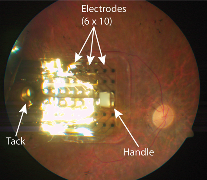
Photograph of an implanted Argus II retinal prosthesis (Second Sight Medical Products, Inc., Sylmar, CA) secured to the retina with a retinal tack. Humayun’s publication describing the clinical trial for this device ranks eighth amongst the most cited articles on pars plana vitrectomy ever published.
Humayun, who serves as director of the USC Dr. Allen and Charlotte Ginsburg Institute for Biomedical Therapeutics and co-director of the USC Roski Eye Institute, reached the top echelon of this list for his contributions to developing a microsurgical instrument system that obviates the need for suturing the eye wall (sclera) after performing retinal surgery. He was also recognized for his pioneering work to establish a retinal implant that partially restores vision to blind patients.
Three out of Humayun’s four top-ranking publications center on a transconjunctival sutureless vitrectomy system (TSV) composed of 25-gauge instruments and accompanying tubes called cannulas through which the instruments are inserted into the eye. Prior to this system, most vitreoretinal surgeons regularly used larger 20-gauge surgical instruments that left behind incisions in the sclera requiring suturing. Their added bulk also traumatized the ocular tissue and resulted in longer recovery times for patients. Humayun and his colleagues Gildo Fujii, MD, and Eugene de Juan, MD, shared their 25-gauge system with the medical community in 2002 and offered surgeons a toolkit that decreased tissue damage, operating times and patient recovery times, all while leaving behind self-sealing incisions. Today, that publication ranks as the fourth most cited of all vitrectomy articles, and their paper discussing their initial experiences using the TSV system ranks sixth. Their follow-up study tracking the outcomes of 140 consecutive cases of 25-gauge transconjunctival surgery for diseases of the back of the eye ranks twelfth.
Humayun’s work appears a fourth time amongst the top twelve articles for his invention and implementation of the Argus II retinal prosthesis, which partially restores vision to blind patients. The device sits inside the eye, attached to the retina, and is implanted via a pars plana vitrectomy approach.
In 2012, Humayun and his colleagues published clinical trial results demonstrating that the device enabled previously blind patients to locate objects, sense motion and detect the orientation of angled, moving lines on a computer screen. The Argus II prosthesis was a groundbreaking invention that ranks eighth amongst the most cited vitrectomy publications, and its development earned Humayun the National Medal of Technology & Innovation from President Obama in 2016.
“It is great to know that our publications on the TSV system and the Argus II prosthesis are some of the most cited articles in the field of ophthalmology,” Humayun says. “I feel privileged to have worked with such excellent collaborators to impact the lives of countless patients, and I look forward to new opportunities to continue driving innovation in our field.”
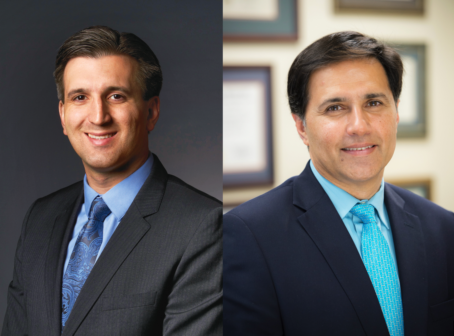
Vitreoretinal surgeons Amir Kashani, MD, PhD (left) and Mark Humayun, MD, PhD (right) pioneered a new surgical procedure to treat dry age-related macular degeneration.
Dry age-related macular degeneration (dry AMD) poses a significant clinical challenge. It is one of the leading causes of progressive blindness, robbing millions of people over the age of 65 of their central vision, and it often hinders patients’ abilities to read books, drive and discern the faces of their loved ones. Although vitamin-based supplements may slow progression, no treatments currently exist.
A team of physicians and scientists at the USC Dr. Allen and Charlotte Ginsburg Institute for Biomedical Therapeutics (Ginsburg Institute) saw in this situation an opportunity to innovate and pioneer a novel treatment approach for dry AMD patients. Theirs has been a feat of scientific and surgical prowess, and over a decade of their diligence and ingenuity has resulted in what may become the first FDA-approved treatment to transform the prospects of regaining vision for millions of patients.
The Ginsburg Institute team, led by vitreoretinal surgeons Amir Kashani, MD, PhD, associate professor of ophthalmology at the Keck School of Medicine, and Mark Humayun, MD, PhD, director of the Ginsburg Institute and co-director of the USC Roski Eye Institute, developed a stem cell-based retinal implant and accompanying surgical procedure to help restore vision to dry AMD patients. Their innovative approach and insights from their phase 1/2a clinical trial are described in the latest print issue of the American Academy of Ophthalmology’s journal Ophthalmology Retina.
DESIGNING THE IMPLANT
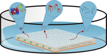
The team accomplished a remarkable multi-part feat that required inventiveness at every turn, starting with designing the novel retinal implant. Dry AMD causes a single layer of cells in the retina called the retinal pigment epithelium (RPE) to deteriorate. The Ginsburg Institute team decided to utilize stem cells to grow RPE tissue in the lab, with the ultimate goal of implanting those cells in patients’ eyes to slow or reverse the damage. Other scientists had attempted to inject stem cell-derived RPE cells into the retina, but had trouble getting the cells to evenly disperse; the Ginsburg Institute scientists instead created a thin membrane made of parylene on which to grow the cells in a single, even layer. Once they had created this RPE layer, the next challenge was to successfully implant it in the eye.
“In practice, being able to get underneath the retina, which is only about a quarter of a millimeter thick, to physically replace the RPE cell layer is a challenging task,” explains Kashani, who is lead author of the publication. “Normally we don’t operate underneath the retina. It’s a place you generally try to avoid during surgery, so that has been a very novel, challenging aspect of delivering these stem cells.”
There are very few tools for performing surgery within the subretinal space. Most available tools were designed 30 to 40 years ago, are relatively bulky and are generally meant to remove scar tissue or other lesions rather than insert anything into the subretinal space. The Ginsburg Institute team decided that the most promising option was to start fresh and design a brand-new tool to fit their purpose.
ENGINEERING THE TOOL
This new tool had to fit a number of criteria: it needed to be made of completely non-toxic materials so as not to harm patients, its design had to be easily reproducible, and it had to be small enough—on a scale of millimeters—to perform minimally invasive surgery inside the eye but large enough to prevent crushing the tissue implant it was meant to deliver.
The surgeons worked with materials and design engineers at the Ginsburg Institute to create single-use forceps with an internal compartment to encapsulate the implant and a roller-style thumbwheel to deploy it. The implant itself is shaped much like a champagne bottle, and the forceps grab onto the narrow end. Rolling the implant into the device’s compartment causes it to fold into a curved shape, and the surgeon can ultimately release it to lay flat inside the eye.
PIONEERING THE SURGICAL TECHNIQUE
With the new instrument and implant came an entirely novel surgical approach. Kashani and Humayun needed to figure out how to create space for the implant in the location of geographic atrophy, which is what doctors call the area of tissue degeneration. To do so, the surgeons decided to create an artificial retinal detachment using a technique called bleb formation, in which a small pocket of space is formed under the retina. “Normally we treat retinal detachments, we don’t make them. In this particular case, we had to create a very well-controlled retinal detachment within an area of scar tissue that is very adherent to the surroundings,” says Kashani. “The challenge was to separate it without damaging the retina.”
In pre-clinical models, creating a bleb alone proved insufficient; the surgeons had to innovate again and ultimately used water pressure to dissect one cell layer from another in a process called targeted hydrodissection. To monitor progress during surgery and prevent complications, the team utilized an advanced imaging technique called optical coherence tomography (OCT) to visualize the dissection at the cellular level. “One part of our job was to make this a very doable surgery and I think we have achieved that with this study,” Kashani says.
“Without tools like OCT, it would be very difficult to visualize the damage we need to treat,” Kashani explains. He emphasizes that in addition to using OCT intraoperatively, he sees a promising role for the technology to be used in earlier-stage AMD patients to monitor the progression of their geographic atrophy. “It’s not a standard of practice to use OCT and other diagnostic methods to detect early and subtle disease changes, but that may prove to be really important for classifying disease and treating it in the future.”
Kashani adds that one of the most rewarding aspects of the entire clinical trial process has been working with his patients and witnessing their commitment to making this translation from research lab to clinical practice possible. “None of this is happening by magic. Patients are volunteering, and they’re taking a chance for the sake of advancing medicine and potentially helping countless other patients down the road. We always appreciate that effort and we thank the patients and their families, too.”
––
This phase 1/2a trial was supported in large part by a $3.73 million grant from the California Institute for Regenerative Medicine (CIRM). Other authors on the study include Jeremy Uang, BS, of the USC Roski Eye Institute, USC Ginsburg Institute for Biomedical Therapeutics and Department of Ophthalmology, Keck School of Medicine of USC; Melissa Mert, MS, of the Southern California Clinical and Translational Science Institute and USC Department of Preventive Medicine (Biostatistics); Firas Rahhal, MD, of the Retina-Vitreous Associates Medical Group; Clement Chan, MD, of the Southern California Desert Retina Consultants, Palm Desert; Robert L. Avery, MD, of the California Retinal Consultants, Santa Barbara; Pravin Dugel, MD of the Retinal Consultants of Arizona, Phoenix; Sanford Chen, MD, of Orange County Retina, Santa Ana; Jane Lebkowski, PhD, of Regenerative Patch Technologies LLC; Dennis O. Clegg, PhD, of the center for Stem Cell Biology and Engineering, University of California; and David R. Hinton, MD, of the Department of Pathology, Keck School of Medicine of USC.
The National Science Foundation recently awarded an Emerging Frontiers in Research and Innovation (EFRI) grant of $1,999,999 to Mark Humayun, MD, PhD, director of the USC Dr. Allen and Charlotte Ginsburg Institute for Biomedical Therapeutics and co-director of the USC Roski Eye Institute. The grant will support an innovative and multidisciplinary project combining engineering and molecular biology to slow down or potentially reverse retinal degeneration. The USC Ginsburg Institute researchers are investigating how electrical stimulation can be used to turn off genes that cause retinal disease while activating “neuroprotective” genes –– named for their abilities to promote healing in the retina, which is part of the central nervous system.
THE CONCEPT
The idea stemmed from a key observation Humayun and his colleagues made while examining patients with severe retinal degeneration who had received Argus II retinal implants, co-invented by Humayun. The Argus II system uses electrical stimulation to transmit visual information from a small camera mounted on the patient’s glasses to the retinal implant, allowing blind patients to regain some eyesight. In patients who received an implant in one eye, Humayun and his team noticed that the area of the retina surrounding the implant was healthier than areas further from the implant or compared to the same regions of the opposite eye. This led them to hypothesize that the electrical stimulation from the Argus prosthesis was somehow preserving neurons within the retina.
They attributed this phenomenon to epigenetic changes, or modifications that happen across the genome to change gene activity. To understand epigenetic changes, think of sports teams. Not all players need to be in the game at once, so the coach decides who plays and who sits on the bench. Similarly, humans have approximately 21,000 protein-coding genes at our disposal, and epigenetic changes act as genomic ‘coaches’ to regulate which genes are active and which sit out at a given time in one’s life. In the case of retinal degeneration, this epigenetic regulation gets thrown off, causing inactivation of genes beneficial to healthy cell function and activation of those that can lead to dysfunction and cell death. USC Ginsburg Institute scientists were among the first researchers to propose that electromagnetic stimulation could be used in patients to alter this epigenetic regulation in a way that helped promote retinal healing.
This ambitious goal would require a feat of bioengineering, combined with in-depth understanding of gene regulation. Humayun, an ophthalmologist and biomedical engineer, teamed up with electrical engineer Gianluca Lazzi, PhD, MBA, translational genomics expert Bodour Salhia, PhD, and a group of scientists at the Northwestern University Center for Physical Genomics and Engineering. Together, the multidisciplinary team is developing a non-invasive, wearable device to electrically stimulate the eye in a safe, controlled way to modify gene expression in the retina and slow or reverse degenerative disease.
USC Ginsburg Institute researchers are developing a contact lens that uses electrical stimulation to heal diseased retinas. (Image: Shutterstock)
TECHNICAL APPROACH
Humayun and Lazzi are working on developing what they call an “e-lens,” which generates electromagnetic fields and can be worn much like a contact lens throughout the night. Now, anyone with contacts has likely been told not to wear them to sleep, because the lenses prevent oxygen from reaching the cornea and, coupled with a closed eyelid, can cause damage due to oxygen deprivation. The e-lens avoids that issue because the team designed it in the shape of a ring that sits around the perimeter of the cornea rather than covering it, making the device safe to wear overnight. Electromagnetic fields from the e-lens would then transmit across the short distance from the cornea to the retina while patients sleep. The e-lens is still in development, but the researchers are working on refining the device and ideally creating inexpensive, single-use coils safe for regular use.
Meanwhile, Salhia is applying her translational genomics expertise to characterize the types of epigenetic changes that electromagnetic fields induce, while working on answering why these changes have a net positive effect on the retina. The team initially identified approximately 3,000 genes that appear to be activated or inactivated due to electrical stimulation, so the goal now is to figure out which of these epigenetic changes are most likely causing the neuroprotective effects.
Crucially, the team has to determine the ideal schedule for retinal stimulation in a theoretical patient. In preliminary studies, the team has identified three groups of genetic responses to stimulation that occur at different speeds: quick and transient genetic changes that show up immediately but are unnoticeable after a few days, quick and sustained changes, and delayed genetic changes. The research team at Northwestern is currently using a computer model of the retina to better track these changes both spatially and over time. Using this information, the team will determine the ideal frequency and duration of time that patients would need to wear the e-lens to benefit from long-lasting neuroprotection.
FUTURE IMPACT
Ideally, this non-invasive treatment could benefit patients with conditions such as retinitis pigmentosa (RP), primary open-angle glaucoma (POAG) and age-related macular degeneration (AMD). The technology could preemptively slow disease in early-stage patients, or it could serve as an adjunct treatment to complement drug or surgical interventions for patients with late-stage retinal degeneration.
Most cases of retinal degeneration aren’t due to just one defective gene, so the fact that this technique affects an entire gamut of genes and leads to coordinated activation of beneficial changes makes it especially promising. The researchers’ findings that electrical stimulation can inhibit cell death and promote healing likely have far-reaching potential to benefit patients with other neurodegenerative diseases and neural injuries like strokes or closed-head injuries.
“That’s the beauty of translational genomics –– it transcends disciplines. You can apply epigenomics and genomics technologies to any disease or system you want to study,” Salhia says. “It grows our world a little bit, because we have technology that we can share with others,” she continues, adding that these applications have a great deal of potential to impact human health moving forward.
“This is a very left field approach that is the result of the efforts of a highly interdisciplinary team of scientists working together,” Salhia continues, adding that one of the most rewarding parts of this project has been collaborating with researchers from widely different backgrounds and using the project as an opportunity to teach each other about their respective disciplines.
“It’s very sci-fi, and we’re really just starting at the beginning,” Salhia continues. “This is just the tip of the iceberg, and this grant is going to allow us to really expand our efforts.”
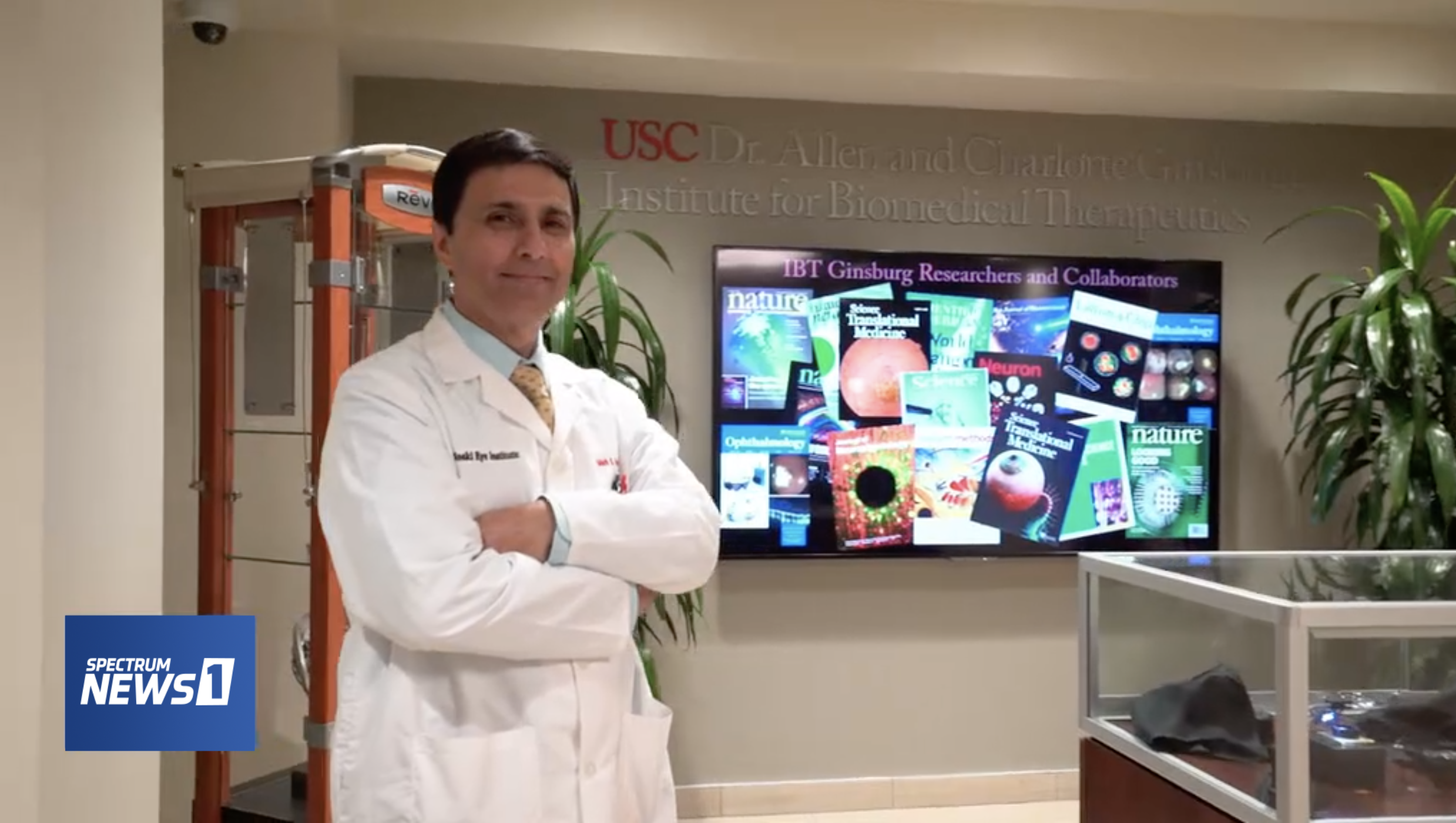
Mark Humayun, MD, PhD, discusses his scientific achievements and inspiration to cure blindness on LA Stories with Giselle Fernandez (Image: Spectrum News).
Mark Humayun, MD, PhD, was featured in an episode of LA Stories with Giselle Fernandez, an Emmy Award-winning series on Spectrum News 1 dedicated to highlighting the “change agents” of Southern California who are shaping the future and imparting lasting changes on our community. Humayun, who serves as director of the USC Dr. Allen and Charlotte Ginsburg Institute for Biomedical Therapeutics and co-director of the USC Roski Eye Institute, was recognized for his pioneering work to treat blindness. On the episode, he shared his family history of practicing medicine, his inspiration to pursue a cure for blindness and his scientific achievements that impact a growing number of patients’ lives each day.
Humayun comes from a long lineage of physicians, but it was his grandmother’s progressive vision loss that spurred his interest in ophthalmology. He had originally planned on becoming a neurosurgeon, but watching his grandmother suffer alerted him to the urgency of curing blindness and he has since dedicated his career to that goal. If his late grandmother could see him today, she would be proud of his tireless commitment to advance the treatment for blindness, from developing the world’s first “bionic eye” to pioneering a stem-cell based treatment to revive the health of patients’ otherwise-degenerating retinas.
Humayun is widely recognized for developing the Argus II retinal implant, which uses electrical stimulation from a computer chip to help blind patients regain some of their eyesight. In 2016, he was awarded the National Medal of Technology and Innovation from President Barack Obama for development of the Argus II retinal implant and his profound and lasting contributions to advancing the biomedical sciences.
Humayun says the goal of developing a bionic eye initially seemed impossible to achieve, but memories of his grandmother’s struggle pushed him to stay dedicated despite the many challenges his team faced. Flash forward to today, and the implants have helped over 300 blind patients around the world partially regain their eyesight and enjoy an improved quality of life. “This idea of putting a computer chip in the eye to restore sight was truly science fiction, but we made it science fact,” Humayun says.
In addition to creating bionic eyes, Humayun and his team at the USC Ginsburg Institute have recently developed a stem-cell based retinal implant to treat age-related macular degeneration, the leading cause of blindness in the United States. Humayun hopes to further enhance the existing implants and make them available to more people. His team is currently integrating features such as infrared technology to help partially blind patients notice and avoid objects like a hot stovetop that could burn them. Such features could be seen as gifting patients with “superhuman” abilities, but Humayun quotes one of his patients who said that rather than making him feel like a cyborg, the bionic eye helps him feel more human because it allows him to interact with his surroundings more like everyone else.
Proof of Humayun’s life-changing work lies in his patients’ testimonies. Terry Byland, the only person in the world to have Argus implants in both of his eyes, was able to see the outline of his teenage son for the first time thanks to the devices. Anna Kuehl, one of the first patients to receive the stem-cell based retinal implant for macular degeneration, has regained enough of her eyesight to see her husband’s face again. Witnessing patients like Byland and Kuehl regain many of the lost joys in their lives inspires Humayun and often makes him think back to his own grandmother to appreciate how far treatment for blindness has come –– and how much further it has yet to go.
“Having that understanding of what my grandmother went through and this understanding of what we have been able to do, I’m able to talk to [patients] and provide them with hope,” Humayun says, and his commitment to enhancing the treatment of blindness despite its challenges is evident. “You just have to say, I’m going to solve this problem, I don’t know how long it’s going to take, but however long it takes, we’ll give it the time because it is worth it.”
To watch Humayun’s complete interview on LA Stories with Giselle Fernandez, click here.
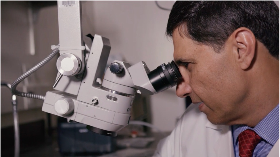
USC Ginsburg Institute researchers were featured for their innovations to restore sight in a recent episode of Voice of America’s “VOA/TEK” (Image: still from “VOA/TEK” episode)
Mark Humayun, MD, PhD and Amir Kashani, MD, PhD were featured on Voice of America’s “VOA/TEK,” a news program dedicated to highlighting the most cutting-edge technologies and medical breakthroughs around the world. The episode details the strides Humayun and his team have made in their mission to restore sight, the lasting impact of their innovations on patients’ lives and the research that continues to evolve at the Dr. Allen and Charlotte Ginsburg Institute for Biomedical Therapeutics.
The episode opened with a feature on Anna Kuehl, a patient who regained her eyesight after receiving a stem cell-based retinal implant as part of a study at the USC Roski Eye Institute. Kuehl had suffered from progressive vision loss due to age-related macular degeneration (AMD), a disease that affects approximately 11 million Americans. She was diagnosed with the dry form of AMD, which causes a layer of cells called the retinal pigment epithelium (RPE) to die off. RPE cells are responsible for nurturing photoreceptor cells in the eye that enable proper eyesight. The loss of functional RPE and photoreceptor cells gradually blinds patients like Kuehl and, historically, there was little doctors could do to help.
Kuehl was one of a handful of people who participated in a clinical trial led by Kashani at the USC Roski Eye Institute, which used stem cells to grow new RPE cells for dry AMD patients. Kashani and Humayun implanted the brand-new cells, grown in a single layer on a biocompatible membrane, to replace the dying RPE cells in Kuehl’s retina.
“When they’re just-formed RPE cells, they’re as young and vibrant as they can get,” Humayun explains in the episode. “They’re incredibly resistant to stressors in the environment, which otherwise would kill older RPE cells. These vibrant, tough cells that we put in Anna’s eye were put in a very destructive environment –– of course, because that environment had killed her cells –– so the question was, would these cells survive?”
The cells did better than survive. They restored Kuehl’s eyesight to the point that she could clearly see the faces of her loved ones once again. During her interview with “VOA/TEK,” Kuehl recalls the moment she first realized she was able see whole faces after the surgery, while she was watching TV: “I jumped back! I was so excited,” she recounts with a laugh.
Kuehl is one of hundreds of patients who have benefitted from the innovations Humayun and his colleagues continue to develop at the USC Ginsburg Institute. Another such patient is Terry Byland, the only person in the world to have two “bionic eyes.”
Byland has a genetic form of blindness called retinitis pigmentosa (RP), which initially causes tunnel vision and eventually leads to complete vision loss. The disease left him in total darkness for 26 years until he received an Argus I and later an Argus II implant, which feed visual information from a camera to the optic nerve to restore some eyesight. The tiny implants are placed inside the eye along the retina and allow patients like Byland to perceive some light and basic motions. The implants offer Byland practical benefits, such as noticing a car in front of him, as well as invaluable gifts, such as the opportunity to see the outline of his now 30-year-old son –– a sight Byland had last witnessed when the boy was only five.
There’s no doubt these implants have brought immeasurable benefits to patients’ lives, but they didn’t arrive at the clinic without challenges. Humayun describes creating the implant as “skiing uphill” because of the immense difficulty of the entire process, from determining what kind of electrical stimulation would produce vision to figuring out how to mount an implant in the tiny, delicate retina. It took an impressive feat of ingenuity and perseverance to develop the implants in the first place, and Humayun is determined to keep making progress. Recently, Argus patients in Korea have begun reporting that they can see the top letter on an eye chart –– a development Humayun finds promising and exciting.
In addition to enhancing the previously described implants, a brain implant based on the Argus platform is also in the works. This device, called Orion, sits directly on the surface of the brain to stimulate a region called the visual cortex. It is intended for patients who have damage to the nerve connecting the eye to the rest of the brain. A patient wears glasses with a tiny camera mounted on the bridge, similar to the glasses and camera used with the Argus retinal implant. As the wearer turns his or her head to take in the surroundings, the camera, instead of transmitting visual information to the implant on the retina, now directly transfers it to the brain implant.
Currently, the implants only sit on one of the brain’s hemispheres and transmit signals through 60 electrodes –– a small quantity compared to the number of signals the estimated 140 million neurons in one’s visual cortex can carry. Second Sight, the company that manufactured the Argus and Orion implants, hopes to eventually increase the number of electrodes, implant devices in both hemispheres and add features like heat vision to help patients identify their family, friends and pets. Six patients have received the Orion implants so far, and they are working with researchers and doctors to retrain their brains to adapt to this new modality of visual information.
“It’s been exciting because every time I come here, I get to see something,” says Benjamin Spencer, one of the Orion implant recipients, during one of his post-implantation testing visits. “It may not be full vision yet, but it’s something, and for someone who hasn’t seen anything in 25 ½ years, that is a huge accomplishment.”
Mark Humayun, MD, PhD, is a co-inventor of the Argus implant series. He is a minority equity owner in Second Sight Medical Products, Inc. and receives royalty payment.
The technology to produce the stem cell–based retinal implant is exclusively licensed to Regenerative Patch Technologies LLC from the University of Southern California, the California Institute of Technology and the University of California, Santa Barbara. Humayun has an equity interest in and is a consultant for Regenerative Patch Technologies LLC.
Researchers at the University of Southern California have developed an economic model to quantify the benefits of treatment for wet age-related macular degeneration (wAMD), the leading cause of blindness in western countries. Their work signals a step forward in the way ophthalmologists audit their practices to define the worth of modern treatments both to patients and society at large.
The study, led by Karen Mulligan, PhD of the Sol Price School of Public Policy and the USC Schaeffer Center for Health Policy & Economics, Seth Seabury, PhD, director of the Keck-Schaeffer Initiative for Population Health Policy and associate professor in the USC School of Pharmacy, and Mark Humayun, MD, PhD, director of the Dr. Allen and Charlotte Ginsburg Institute for Biomedical Therapeutics and co-director of the USC Roski Eye Institute, was published on November 14th in the Journal of the American Medical Association (JAMA). This research represents the first analysis to quantitatively compare the economic benefits of therapeutic injections for wAMD to their costs and assess the value they bring to society. The researchers’ model showed that treating wAMD can generate $5.1 to $8.2 billion in patient benefit and $0.9 to $3 billion in societal benefit in three years based on the value of vision gains alone –– not including other potential benefits such as reductions in medical expenditure or caregiver burden. These numbers are poised to climb even higher if future innovations in health care can boost treatment initiation and adherence rates.
QUANTIFYING THERAPEUTIC VALUE
Wet age-related macular degeneration is a progressive form of blindness caused by the abnormal growth of blood vessels underneath the retina. A molecule called vascular endothelial growth factor (VEGF) drives this blood vessel formation, and doctors can treat the disease with a targeted therapy against that molecule to block blood vessel growth and restore patients’ eyesight for up to five years. Anti-VEGF therapy is largely successful when injected into the eye regularly, yet many people with wAMD never initiate therapy and studies based on Medicare beneficiaries have shown that approximately 53-58% of wAMD patients receiving treatment discontinue within the first year.1,2 Cost, fear of discomfort related to the injection process and the time-consuming nature of follow-up care all contribute to this problem, prompting many doctors to modify treatment schedules to minimize the number of injections a patient must receive without drastically compromising effectiveness.
Finding the balance between providing enough treatment and minimizing the burden on patients can be tricky, and until now it was made even harder by the fact that no one had previously quantified the benefits that anti-VEGF therapy provides for patients and society in exchange for the costs associated with treatment. In an era when health care expenditure is growing at an unsustainable rate and policymakers are looking to cut costs where possible, this situation highlights the importance of quantifying which therapies are most worth their investments.
Other specialties have started a movement of quantifying treatment value and comparing benefits to costs, prompting Humayun to help pioneer this paradigm shift in ophthalmology by partnering with public policy experts like Mulligan and Seabury to audit the field’s own practices.
AN IMPRESSIVE RETURN ON INVESTMENT
Humayun and his team started by combing through scientific literature and accruing model data of wAMD patients treated with anti-VEGF therapy. They translated the patients’ changes in visual acuity over time to “quality-adjusted life years,” which is an economic measurement that reflects both quantity and quality of years lived and could be used as a variable in the team’s economic model.
“Our model assumes a person with perfect, 20/20 vision has a quality of life that is valued at $150,000 for a single year,” Mulligan explains, referencing a commonly used value in health economics. “For a person who is diagnosed with wAMD, their vision is worse –– reflecting that, their quality of life is valued at about $105,000 for a single year,” she continues. “However, if they are treated with anti-VEGFs, their vision gains translate into nearly $11,000 in value after one year. Put another way, a patient with wAMD would be willing to pay $11,000 for the improved quality of life associated with better vision from the first year of anti-VEGF treatments,” she summarizes.
The model reflected multiple treatment scenarios, all compared back to a “No Treatment” control group: patients in the “Less Frequent Injections” group who received approximately 8 injections per year, those in the “More Frequent Injections” group who received on average 10.5 injections per year (a number closer to drug-label treatment suggestions), and several different treatment innovation scenarios to reflect the gains that might occur if more patients initiate and follow through with treatment.
After creating this model, the team used it to simulate a range of possible patient outcomes based on each scenario and compared the benefits of each outcome to its associated costs.
Their findings overwhelmingly point to the vast economic benefits anti-VEGF therapies bring to patients and society as a whole, even in light of the drugs’ relatively high costs. For example, a single patient receiving less frequent injections for a five-year period can benefit from approximately $49,558 in economic gains, and a patient receiving more frequent injections during that time could witness $84,873 worth in benefits from improved quality of life.
The study further bolsters the case for increasing the number of injections per year, providing that patients respond well and experience meaningful vision gains from treatment. Their analysis shows that receiving less frequent injections accrues $873 million in population-wide value over three years while more frequent injections add $2.1 billion to societal value in three years even after accounting for the higher costs of performing more injections. Importantly, the study points out that innovations to improve treatment adherence could generate an additional $1.2 to $3.7 billion in patient benefit and $59 million to $1.3 billion in societal value compared to current treatment scenarios, highlighting the fact that when patients follow through with necessary treatment, both individuals and society as a whole can reap the rewards.
The researchers also demonstrated that if 100% of patients who needed therapy actually initiated treatment and only 6% dropped out per year (the dropout rate in clinical trial data), patients could benefit 42% more compared to simply improving treatment adherence and 89% more than they benefit from the current “Less Frequent Injections” model. In fact, in this “best case” scenario reflecting high rates of both treatment uptake and adherence, the three-year benefits could reach as high as $9.7-$15.0 billion depending on whether patients receive less or more frequent injections respectively.
Finally, the team pointed out that while politicians often scorn the high costs of anti-VEGF treatments (in 2015, Medicare Part B payed out $3.0 billion between just two anti-VEGF drugs), the team showed that relying more on a less expensive type of anti-VEGF drug known as bevacizumab could reduce costs for the full population by as much as $1.8-$2.2 billion over three years. Findings like these point to the importance of using economics to quantify the value of different therapies and auditing common practices in ophthalmology to optimize the way doctors approach treatment in a way that benefits patients and society at large. Escort Paris
Curtis LH, Hammill BG, Qualls LG, et al. Treatment patterns for neovascular age-related macular degeneration: analysis of 284 380 medicare beneficiaries. Am J Ophthalmol. 2012;153(6):1116-1124.e1. doi:10.1016/j.ajo.2011.11.032
Lad EM, Hammill BG, Qualls LG, Wang F, Cousins SW, Curtis LH. Anti-VEGF Treatment Patterns for Neovascular Age-Related Macular Degeneration Among Medicare Beneficiaries. American Journal of Ophthalmology. 2014;158(3):537-543.e2. doi:10.1016/j.ajo.2014.05.014
The California Institute for Regenerative Medicine (CIRM) recently awarded $3.73 million to Mark Humayun, MD, PhD, who serves as director of the USC Dr. Allen and Charlotte Ginsburg Institute for Biomedical Therapeutics and co-director of the USC Roski Eye Institute. The grant is part of CIRM’s translational research program to propel California forward as a hub of regenerative medicine breakthroughs. The funding will support researchers at the USC Ginsburg Institute as they develop a new treatment for dry age-related macular degeneration (dry AMD) –– a disease that has historically been considered difficult to treat.
This injection is not a man-made drug, but is instead composed of molecules produced by healthy retinal cells cultured in the laboratory. The researchers have already demonstrated that an intraocular injection of these molecules stimulates healing and slows down retinal degeneration in pre-clinical studies. The team’s prestigious CIRM grant is intended to accelerate the project’s transition to the clinical trial stage, thanks to its potential to benefit society by filling an unmet medical need.
One innovation inspires another
Approximately two million Americans currently live with advanced forms of AMD, and more than seven million deal with early-stage symptoms of the disease. Of those cases, 85-90% are the dry form of AMD. The condition is characterized by the progressive deterioration of the macula, or the region of the retina responsible for the clearest, most focused vision in the center of one’s field of view. A single layer of cells under the macula called the retinal pigment epithelium (RPE) is responsible for nurturing the eye’s photoreceptor cells, which lie directly on top of the RPE and perceive light.
As dry AMD progresses, patients perceive a dark spot obscuring the center of their vision. (Image: National Eye Institute, National Institutes of Health)
As the macula deteriorates and RPE cells die off, the photoreceptor cells necessary for vision gradually follow suit. Soon, patients begin to perceive a dark spot in the center of their visual field that can interfere with anything from reading to recognizing the faces of loved ones. The disease robs patients of both their eyesight and a great deal of their autonomy.
With the help of a CIRM Disease Team grant, a team of USC Ginsburg Institute researchers recently used stem cells to develop a retinal implant composed of a single layer of RPE cells to replace the degenerating part of the macula. Humayun, working alongside Amir Kashani, MD, PhD, surgically placed implants in 15 patients’ retinas. Although the trial was only intended to assess the implant’s safety, the team observed early signs of therapeutic benefit and a few patients even regained some of their eyesight. That project is currently poised to enter a larger clinical trial phase with the goal of becoming the first ever FDA-approved treatment for late-stage dry AMD.
While testing the implants in pre-clinical models, the team made an intriguing observation: the implanted cells had a restorative effect not only in the exact location they were placed, but also on the surrounding retinal tissue.
The researchers attributed this phenomenon to something they call the paracrine effect: cells from the implant produce chemical messages that communicate with surrounding cells –– a process known to biologists as “paracrine signaling” –– which causes the degenerating native cells to behave more like the healthy, newly implanted ones.
USC Ginsburg Institute researchers are culturing healthy retinal cells and harnessing the biological factors they produce to create therapeutic injections and reverse the symptoms of dry AMD. (Image: Kabir Ahluwalia)
The researchers asked themselves a key question: if these biological factors alone could have a restorative effect on a degenerating retina, could the team create an injectable solution containing the factors to complement the implant as an early-stage intervention for dry AMD?
So far, the answer appears to be yes. Cells used in the implants are grown in the lab in a nutritious broth called the media, and as they grow, the molecular factors they produce are released into the media. By harvesting that factor-filled fluid and delivering it as an intraocular injection, the team has already seen success in dramatically slowing down the progression of retinal cell loss in pre-clinical models of retinal degeneration. The team’s long-term goal is to develop a therapeutic injection for early-stage patients to slow disease progression, while patients whose diseases have progressed to the point of blindness can receive implants.
Sights set on a future cure
Now that the researchers know this media holds promise as a therapeutic injection, the next step is to fully characterize its components. While the team has narrowed down the number of potentially therapeutic molecules in the media, no single constituent seems to have a strong effect on its own, meaning the factors may work together in a synergistic way to restore retinal function. Funding from the CIRM grant is meant to expedite the process of characterizing the media and creating a stable, reproducible therapeutic injection for eventual use in humans.
Kabir Ahluwalia, a doctoral student in the USC School of Pharmacy working on the project, explains that this research holds particular promise for those estimated seven million early-stage patients making up the majority of dry AMD cases. Early-stage AMD patients can currently benefit from specific nutritional supplements that slow down disease progression by about 25%, but many patients still need additional therapy. This novel injection of soluble growth factors could potentially serve as an ideal future treatment to prevent vision loss and provide renewed hope for these patients.
Dr. Mark Humayun from the USC Dr. Allen and Charlotte Ginsburg Institute for Biomedical Therapeutics was recently featured in Nature Outlook for his outstanding contributions to advance the treatment of blindness. The article highlighted a handful of the world’s top researchers tackling the problem of retinal degeneration, which is the leading cause of blindness in developed countries.
Image: Nature Outlook
Dr. Humayun’s Argus series implants were showcased amongst the most cutting-edge approaches to restoring eyesight for patients with some functional retinal cells still intact. The Argus II “bionic eye” consists of an electrode array that is implanted on the surface of the retina. The patient wears glasses equipped with a small video camera that transmits signals wirelessly to the implant. The electrodes stimulate the retina, which then communicates those signals to the brain. Over 300 patients have received the Argus II prosthesis and have regained their perception of light patterns, movement and basic shapes.
For patients who have completely lost functionality of the retina, Humayun and his colleagues at Second Sight have a different approach: sending signals from a camera directly to the brain.
The researchers have developed a chip, called Orion, which is surgically implanted on the outer surface of the very back of one’s brain. This region of the brain, called the visual cortex, is responsible for processing and interpreting information from the eyes. Like Argus II, Orion receives signals from a camera mounted on the patient’s glasses, and the brain can then convert those signals into visual information. So far, the chip has been successfully implanted in five patients with limited or no light perception. The trial is still its early stages, but the preliminary results look promising and Humayun hopes the chip will receive FDA approval in a few years.
To read the entire Nature Outlook article, click here.
A team of researchers at the USC Dr. Allen and Charlotte Ginsburg Institute for Biomedical Therapeutics recently developed a pair of augmented reality (AR) glasses to help visually impaired patients navigate their surroundings and perceive depth more clearly.
The glasses were designed to help patients with a degenerative eye disease called retinitis pigmentosa (RP). The condition causes progressive vision loss, particularly on the periphery of one’s vision, and makes it difficult to see in low-light conditions. Patients with RP often experience tunnel vision and have trouble perceiving their 3D environment. Specifically, they struggle to grasp objects and avoid obstacles in their path, and these issues are worse at night.
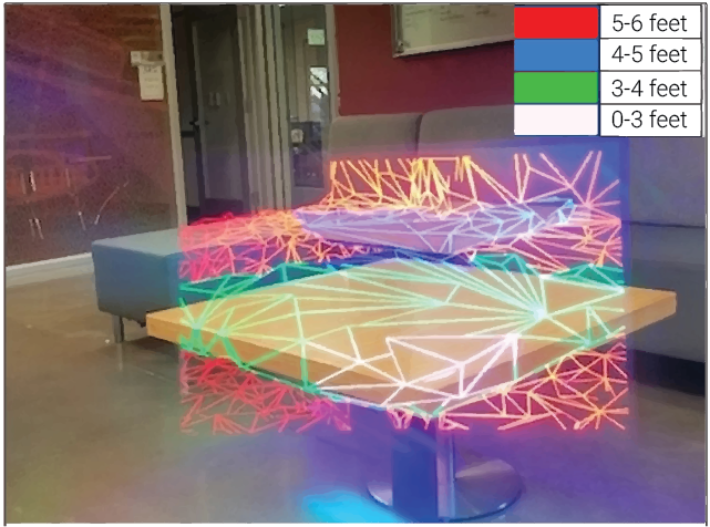
The AR glasses help solve this problem by allowing patients to see a color-coded mesh on top of the objects in their surroundings. The colors correspond to depth, with objects closest to the wearer appearing white, followed by green, blue and eventually red for objects that are furthest away.
The augmented reality glasses allow patients to perceive a color-coded mesh on top of the objects in their surroundings, which helps wearers perceive depth. (Image: Anastasios Nikolas Angelopoulos)
The device, customized by Anastasios Nikolas Angelopoulos and Dr. Mark Humayun, was configured with the user’s experience in mind. Rather than using virtual reality, which completely replaces the wearer’s field of view with an image on a screen, the augmented reality color mesh enhances the wearer’s depth perception while still allowing the patient to see the true color and texture of an object through gaps in the mesh. This is important for patients, because it allows them to interact with the world around them as normally as possible without having to sacrifice any of the perception they still have.
With the help of their colleagues, Drs. Hossein Ameri and Debbie Mitra, the researchers tested the visual aid in subjects with retinitis pigmentosa. The team asked patients to both navigate a simple obstacle course and grasp objects in front of them while they had the glasses on. When using the glasses, RP patients were able to navigate the maze and avoid obstacles 50% better than they could without the visual aid. The grasping task required patients to grab the furthest of four pegs placed in front of them, without hitting any of the closer ones. The glasses improved the patients’ abilities to grasp the furthest peg by 70%, meaning that much of their depth perception was restored thanks to the AR color mesh.
Currently, many patients with RP avoid going out at night and may experience anxiety or fear of losing their independence due to their vision problems. Although the device is still in development, the researchers hope these glasses will eventually help improve quality of life by allowing patients to return to their day-to-day activities safely and with more confidence and independence.
The team published their work in Scientific Reports, an online, open access journal from the publishers of Nature, and the article has been accessed close to 2,000 times. Their publication ranks in the 98th percentile of all similarly aged papers tracked by the data science company Altmetric across all journals, and it ranks 1st of 12 tracked articles of a similar age in Scientific Reports. The researchers’ work was additionally featured in news outlets such as ScienceDaily, and the full list of media coverage is available here.
 Mark Humayun, MD, PhD (Photo credit: USC Roski Eye Institute)