USC research coalition uses computer models to accelerate advancements in vision science

By Alexandra Demetriou

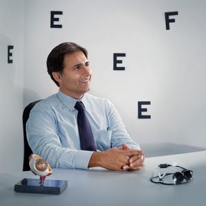
The Argus II retinal prosthesis––often referred to as the world’s first “bionic eye” ––revolutionized the treatment of blindness. Throughout the past decade, this artificial retina, developed by Mark Humayun, MD, PhD and colleagues in collaboration with Second Sight Medical Products, has enabled hundreds of patients around the world with complete retinal blindness to regain partial eyesight.

The Argus II retinal prosthesis enables patients with complete retinal blindness to see the shapes of objects in their surroundings, such as this Fourth of July fireworks show.

The invention currently allows for black-and-white vision and particularly helps patients detect boundaries between objects in their surroundings. Using a combination of patient feedback and laboratory experiments, Humayun and his team at the USC Dr. Allen and Charlotte Ginsburg Institute for Biomedical Therapeutics continue to enhance the visual information retinal prostheses can offer patients.

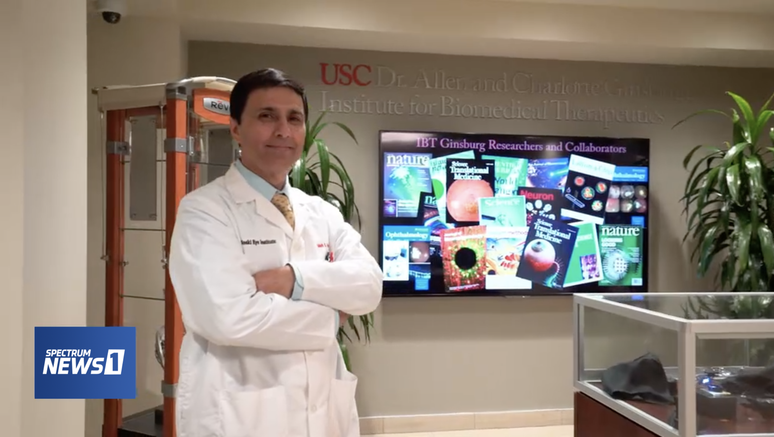
In their latest work, USC Ginsburg Institute scientists, in collaboration with USC Institute for Technology and Medical Systems (ITEMS) engineers, have developed detailed computer models of the retina that enable them to test modifications to the prosthetic system with unprecedented ease and speed. These advances in computational modeling have enabled the team to make profound strides forward in understanding how to refine and build upon the device’s capabilities––with the long-term goal of restoring color vision and more precise visual details to patients with progressive blindness.

A look inside the “bionic eye”

At the most basic level, retinal prostheses work by converting visual information from the environment into electrical impulses that stimulate the otherwise blind retina. When these signals reach the vision center of the brain, known as the visual cortex, patients are able to perceive images and shapes from their surroundings.

The Argus II system involves a retinal implant––or, more specifically, a microelectrode array that sits atop and stimulates the retina––coupled with a pair of glasses outfitted with a camera that transmits signals to the implant. When the implant receives visual cues from the camera, it generates small electrical impulses that stimulate the patient’s retinal cells. This stimulation causes the patient to perceive a flash of light that vision scientists refer to as a phosphene.

Image of the Argus II retinal prosthesis, composed of a microelectrode array with thin projections that electrically stimulate the retina.

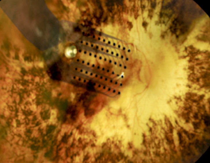
Phosphenes should ideally appear as small, rounded dots when a single electrode stimulates the retina. In practice, however, patients sometimes perceive a dash instead of a dot––and the reason has to do with the way cells intermesh in the retina.

One of these cell types is the retinal ganglion cell, which plays an essential role in sending visual information from the eye to the brain. Retinal ganglion cells have rounded bodies with many thin projections branching off in different directions and one long projection that feeds information to the brain. These projections overlap and intertwine like the branches of trees in a densely wooded forest, so that when an electrode stimulates one of the retinal cells it often activates the projections of neighboring cells as well.

Example of a retinal ganglion cell, composed of a rounded, central body with numerous small projections and one long projection, or axon, that communicates with the brain (red). Vision scientists at the USC Ginsburg Institute are developing techniques to activate the bodies of specific cells without stimulating the projections of nearby cells, which will help sharpen the visual information that retinal prosthetics can offer patients.

When electrical signals from the prosthesis stimulate both cell bodies and projections from nearby cells, patients perceive a dash of light, called an elongated phosphene, instead of a discrete dot. Humayun’s team set out to determine how to prevent elongated phosphenes from forming so patients could enjoy more refined, detailed vision. Doing so required devising an approach to target retinal cell bodies without activating neighboring cells in the process.


Computer models probe deeper into cellular behavior

To solve this challenge, the USC Ginsburg Institute and USC ITEMS teams first developed computational models to recreate different types of retinal ganglion cells and the patterns in which they overlap. Their models accounted for the relevant functional properties of retinal ganglion cells and could therefore respond to stimuli in ways that mimicked how real retinal ganglion cells would respond. Using these model systems, the scientists ran through a gamut of simulations to find a stimulation pattern that activated cell bodies while avoiding the large projections, known as axons, that other cells use to send visual information to the brain.

In one recent study, the team reported their discovery that very short-duration electrical pulses, as brief as 0.1 milliseconds, can selectively target the intended retinal ganglion cells’ bodies without activating their axons. Their models also enabled them to more accurately map out patterns of cellular activation that arise when shorter pulses are used to stimulate the retina. The researchers hope these, and future, findings can help resolve the problem of elongated phosphenes to ultimately offer patients enhanced visual resolution with a retinal prosthesis.

USC Ginsburg Institute and USC ITEMS scientists are also currently using their computational models of the retina to study possible ways of programming additional visual components, such as color, into the Argus II system. A 2020 study of theirs showed that a particular subtype of retinal ganglion cell, called a bistratified cell, is more responsive to high frequency stimulation than one of its counterparts known as a monostratified cell. Understanding these types of slight differences in responses may help the team improve the performance of retinal prosthetics to enable patients to detect color, contrast and edges of objects. Such research is still ongoing, but based on their preliminary findings, the team is hopeful for the ways they can continue enhancing visual perception for the hundreds of retinal prosthesis users who once lived in complete darkness.

Javad Paknahad, a PhD candidate at USC and the lead author on the aforementioned studies, emphasized how the “multi-scale” nature of these models, which account for the properties of single cells as well as the behavior of entire cell populations, makes this approach particularly valuable. He added that using computer models to study the retina allows their team to run experiments more efficiently, which ultimately accelerates scientific progress. “These multi-scale computational models end up saving a lot of time when doing experiments, because as you continue refining the model system and designing the right programs, you can more readily determine the best approaches to solving complex problems,” he said. “This makes computational modeling very powerful.”


Disclosure: Mark Humayun, MD, PhD, is a co-inventor of the Argus implant series.  He is a minority equity owner in Second Sight Medical Products, Inc. and receives royalty payment. 

Disclosures: Regenerative Patch Technologies LLC was founded by Mark Humayun, MD, PhD, and David R. Hinton, MD, from the University of Southern California, and Dennis O. Clegg, PhD, from the University of California, Santa Barbara. The technology to produce the stem cell–based retinal implant is exclusively licensed to Regenerative Patch Technologies LLC from the University of Southern California, the California Institute of Technology and the University of California, Santa Barbara. Humayun has an equity interest in and is a consultant for Regenerative Patch Technologies LLC.

Stanford study ranks Mark Humayun, MD, PhD on world’s top scientists list

By Alexandra Demetriou

Mark Humayun, MD, PhD, was included in a recent analysis out of Stanford University highlighting the world’s leading scientists across 22 fields of research and 176 sub-fields. Within the category of “ophthalmology & optometry,” Humayun ranks among the top 0.2% of his peers.

A recent Stanford study ranked Mark Humayun, MD, PhD, as one of the world's top scientists. (Image credit: Jill Greenberg)
A recent Stanford study ranked Mark Humayun, MD, PhD, as one of the world’s top scientists. (Image credit: Jill Greenberg)

The rankings were calculated based on metrics such as the number of times a scientist’s research has been cited throughout the individual’s career. The analysis also accounted for each person’s h-index score, which attempts to quantify the scientist’s research productivity balanced with the impact that the research has made on the scientific field.

Humayun, a vitreoretinal surgeon and prolific inventor of biomedical devices, is widely known for developing the world’s first artificial retina. In 2016, Humayun received the National Medal of Technology and Innovation from President Barack Obama for developing the Argus II retinal prosthesis, which restores partial vision to patients with total retinal blindness.

Some of Humayun’s other accolades include election to both the National Academy of Medicine (NAM) and National Academy of Engineering (NAE), the 2020 Medal for Innovations in Healthcare Technology from the Institute of Electrical and Electronics Engineers (IEEE), “Inventor of the Year” from R&D Magazine in 2005, and distinction as one of the top 1% of ophthalmologists in U.S. News & World Report.

Humayun currently serves as director of the USC Dr. Allen and Charlotte Ginsburg Institute for Biomedical Therapeutics and co-director of the USC Roski Eye Institute. He also holds the titles of University Professor and Cornelius J. Pings Chair in Biomedical Sciences at USC.

Physician-scientists pioneer new surgical approach to treat progressive blindness

By Alexandra Demetriou

Vitreoretinal surgeons Amir Kashani, MD, PhD (left) and Mark Humayun, MD, PhD (right) pioneered a new surgical procedure to treat dry age-related macular degeneration.

Dry age-related macular degeneration (dry AMD) poses a significant clinical challenge. It is one of the leading causes of progressive blindness, robbing millions of people over the age of 65 of their central vision, and it often hinders patients’ abilities to read books, drive and discern the faces of their loved ones. Although vitamin-based supplements may slow progression, no treatments currently exist.

A team of physicians and scientists at the USC Dr. Allen and Charlotte Ginsburg Institute for Biomedical Therapeutics (Ginsburg Institute) saw in this situation an opportunity to innovate and pioneer a novel treatment approach for dry AMD patients. Theirs has been a feat of scientific and surgical prowess, and over a decade of their diligence and ingenuity has resulted in what may become the first FDA-approved treatment to transform the prospects of regaining vision for millions of patients.

The Ginsburg Institute team, led by vitreoretinal surgeons Amir Kashani, MD, PhD, associate professor of ophthalmology at the Keck School of Medicine, and Mark Humayun, MD, PhD, director of the Ginsburg Institute and co-director of the USC Roski Eye Institute, developed a stem cell-based retinal implant and accompanying surgical procedure to help restore vision to dry AMD patients. Their innovative approach and insights from their phase 1/2a clinical trial are described in the latest print issue of the American Academy of Ophthalmology’s journal Ophthalmology Retina.

DESIGNING THE IMPLANT

The team accomplished a remarkable multi-part feat that required inventiveness at every turn, starting with designing the novel retinal implant. Dry AMD causes a single layer of cells in the retina called the retinal pigment epithelium (RPE) to deteriorate. The Ginsburg Institute team decided to utilize stem cells to grow RPE tissue in the lab, with the ultimate goal of implanting those cells in patients’ eyes to slow or reverse the damage. Other scientists had attempted to inject stem cell-derived RPE cells into the retina, but had trouble getting the cells to evenly disperse; the Ginsburg Institute scientists instead created a thin membrane made of parylene on which to grow the cells in a single, even layer. Once they had created this RPE layer, the next challenge was to successfully implant it in the eye.

“In practice, being able to get underneath the retina, which is only about a quarter of a millimeter thick, to physically replace the RPE cell layer is a challenging task,” explains Kashani, who is lead author of the publication. “Normally we don’t operate underneath the retina. It’s a place you generally try to avoid during surgery, so that has been a very novel, challenging aspect of delivering these stem cells.”

There are very few tools for performing surgery within the subretinal space. Most available tools were designed 30 to 40 years ago, are relatively bulky and are generally meant to remove scar tissue or other lesions rather than insert anything into the subretinal space. The Ginsburg Institute team decided that the most promising option was to start fresh and design a brand-new tool to fit their purpose.

ENGINEERING THE TOOL

This new tool had to fit a number of criteria: it needed to be made of completely non-toxic materials so as not to harm patients, its design had to be easily reproducible, and it had to be small enough—on a scale of millimeters—to perform minimally invasive surgery inside the eye but large enough to prevent crushing the tissue implant it was meant to deliver.

The surgeons worked with materials and design engineers at the Ginsburg Institute to create single-use forceps with an internal compartment to encapsulate the implant and a roller-style thumbwheel to deploy it. The implant itself is shaped much like a champagne bottle, and the forceps grab onto the narrow end. Rolling the implant into the device’s compartment causes it to fold into a curved shape, and the surgeon can ultimately release it to lay flat inside the eye.

PIONEERING THE SURGICAL TECHNIQUE

With the new instrument and implant came an entirely novel surgical approach. Kashani and Humayun needed to figure out how to create space for the implant in the location of geographic atrophy, which is what doctors call the area of tissue degeneration. To do so, the surgeons decided to create an artificial retinal detachment using a technique called bleb formation, in which a small pocket of space is formed under the retina. “Normally we treat retinal detachments, we don’t make them. In this particular case, we had to create a very well-controlled retinal detachment within an area of scar tissue that is very adherent to the surroundings,” says Kashani. “The challenge was to separate it without damaging the retina.”

In pre-clinical models, creating a bleb alone proved insufficient; the surgeons had to innovate again and ultimately used water pressure to dissect one cell layer from another in a process called targeted hydrodissection. To monitor progress during surgery and prevent complications, the team utilized an advanced imaging technique called optical coherence tomography (OCT) to visualize the dissection at the cellular level. “One part of our job was to make this a very doable surgery and I think we have achieved that with this study,” Kashani says.

“Without tools like OCT, it would be very difficult to visualize the damage we need to treat,” Kashani explains. He emphasizes that in addition to using OCT intraoperatively, he sees a promising role for the technology to be used in earlier-stage AMD patients to monitor the progression of their geographic atrophy. “It’s not a standard of practice to use OCT and other diagnostic methods to detect early and subtle disease changes, but that may prove to be really important for classifying disease and treating it in the future.”

Kashani adds that one of the most rewarding aspects of the entire clinical trial process has been working with his patients and witnessing their commitment to making this translation from research lab to clinical practice possible. “None of this is happening by magic. Patients are volunteering, and they’re taking a chance for the sake of advancing medicine and potentially helping countless other patients down the road. We always appreciate that effort and we thank the patients and their families, too.”

––

This phase 1/2a trial was supported in large part by a $3.73 million grant from the California Institute for Regenerative Medicine (CIRM). Other authors on the study include Jeremy Uang, BS, of the USC Roski Eye Institute, USC Ginsburg Institute for Biomedical Therapeutics and Department of Ophthalmology, Keck School of Medicine of USC; Melissa Mert, MS, of the Southern California Clinical and Translational Science Institute and USC Department of Preventive Medicine (Biostatistics); Firas Rahhal, MD, of the Retina-Vitreous Associates Medical Group; Clement Chan, MD, of the Southern California Desert Retina Consultants, Palm Desert; Robert L. Avery, MD, of the California Retinal Consultants, Santa Barbara; Pravin Dugel, MD of the Retinal Consultants of Arizona, Phoenix; Sanford Chen, MD, of Orange County Retina, Santa Ana; Jane Lebkowski, PhD, of Regenerative Patch Technologies LLC; Dennis O. Clegg, PhD, of the center for Stem Cell Biology and Engineering, University of California; and  David R. Hinton, MD, of the Department of Pathology, Keck School of Medicine of USC.

Science fiction to science fact: Mark Humayun, MD, PhD, talks cutting-edge science, family history, and dedication to curing blindness on Spectrum News

By Alexandra Demetriou

Mark Humayun, MD, PhD, discusses his scientific achievements and inspiration to cure blindness on LA Stories with Giselle Fernandez (Image: Spectrum News).

Mark Humayun, MD, PhD, was featured in an episode of LA Stories with Giselle Fernandez, an Emmy Award-winning series on Spectrum News 1 dedicated to highlighting the “change agents” of Southern California who are shaping the future and imparting lasting changes on our community. Humayun, who serves as director of the USC Dr. Allen and Charlotte Ginsburg Institute for Biomedical Therapeutics and co-director of the USC Roski Eye Institute, was recognized for his pioneering work to treat blindness. On the episode, he shared his family history of practicing medicine, his inspiration to pursue a cure for blindness and his scientific achievements that impact a growing number of patients’ lives each day.

Humayun comes from a long lineage of physicians, but it was his grandmother’s progressive vision loss that spurred his interest in ophthalmology. He had originally planned on becoming a neurosurgeon, but watching his grandmother suffer alerted him to the urgency of curing blindness and he has since dedicated his career to that goal. If his late grandmother could see him today, she would be proud of his tireless commitment to advance the treatment for blindness, from developing the world’s first “bionic eye” to pioneering a stem-cell based treatment to revive the health of patients’ otherwise-degenerating retinas.

Humayun is widely recognized for developing the Argus II retinal implant, which uses electrical stimulation from a computer chip to help blind patients regain some of their eyesight. In 2016, he was awarded the National Medal of Technology and Innovation from President Barack Obama for development of the Argus II retinal implant and his profound and lasting contributions to advancing the biomedical sciences.

Humayun says the goal of developing a bionic eye initially seemed impossible to achieve, but memories of his grandmother’s struggle pushed him to stay dedicated despite the many challenges his team faced. Flash forward to today, and the implants have helped over 300 blind patients around the world partially regain their eyesight and enjoy an improved quality of life. “This idea of putting a computer chip in the eye to restore sight was truly science fiction, but we made it science fact,” Humayun says.

In addition to creating bionic eyes, Humayun and his team at the USC Ginsburg Institute have recently developed a stem-cell based retinal implant to treat age-related macular degeneration, the leading cause of blindness in the United States. Humayun hopes to further enhance the existing implants and make them available to more people. His team is currently integrating features such as infrared technology to help partially blind patients notice and avoid objects like a hot stovetop that could burn them. Such features could be seen as gifting patients with “superhuman” abilities, but Humayun quotes one of his patients who said that rather than making him feel like a cyborg, the bionic eye helps him feel more human because it allows him to interact with his surroundings more like everyone else.

Proof of Humayun’s life-changing work lies in his patients’ testimonies. Terry Byland, the only person in the world to have Argus implants in both of his eyes, was able to see the outline of his teenage son for the first time thanks to the devices. Anna Kuehl, one of the first patients to receive the stem-cell based retinal implant for macular degeneration, has regained enough of her eyesight to see her husband’s face again. Witnessing patients like Byland and Kuehl regain many of the lost joys in their lives inspires Humayun and often makes him think back to his own grandmother to appreciate how far treatment for blindness has come –– and how much further it has yet to go.

“Having that understanding of what my grandmother went through and this understanding of what we have been able to do, I’m able to talk to [patients] and provide them with hope,” Humayun says, and his commitment to enhancing the treatment of blindness despite its challenges is evident. “You just have to say, I’m going to solve this problem, I don’t know how long it’s going to take, but however long it takes, we’ll give it the time because it is worth it.”

To watch Humayun’s complete interview on LA Stories with Giselle Fernandez, click here.

Mark Humayun, MD, PhD and Amir Kashani, MD, PhD featured in Voice of America “VOA/TEK” episode highlighting their mission to restore sight

By Alexandra Demetriou

Anna Kuehl, a USC Roski Eye Institute patient featured on Voice of America's
USC Ginsburg Institute researchers were featured for their innovations to restore sight in a recent episode of Voice of America’s “VOA/TEK” (Image: still from “VOA/TEK” episode)

 Mark Humayun, MD, PhD and Amir Kashani, MD, PhD were featured on Voice of America’s “VOA/TEK,” a news program dedicated to highlighting the most cutting-edge technologies and medical breakthroughs around the world. The episode details the strides Humayun and his team have made in their mission to restore sight, the lasting impact of their innovations on patients’ lives and the research that continues to evolve at the Dr. Allen and Charlotte Ginsburg Institute for Biomedical Therapeutics.

The episode opened with a feature on Anna Kuehl, a patient who regained her eyesight after receiving a stem cell-based retinal implant as part of a study at the USC Roski Eye Institute. Kuehl had suffered from progressive vision loss due to age-related macular degeneration (AMD), a disease that affects approximately 11 million Americans. She was diagnosed with the dry form of AMD, which causes a layer of cells called the retinal pigment epithelium (RPE) to die off. RPE cells are responsible for nurturing photoreceptor cells in the eye that enable proper eyesight. The loss of functional RPE and photoreceptor cells gradually blinds patients like Kuehl and, historically, there was little doctors could do to help.

Kuehl was one of a handful of people who participated in a clinical trial led by Kashani at the USC Roski Eye Institute, which used stem cells to grow new RPE cells for dry AMD patients. Kashani and Humayun implanted the brand-new cells, grown in a single layer on a biocompatible membrane, to replace the dying RPE cells in Kuehl’s retina.

“When they’re just-formed RPE cells, they’re as young and vibrant as they can get,” Humayun explains in the episode. “They’re incredibly resistant to stressors in the environment, which otherwise would kill older RPE cells. These vibrant, tough cells that we put in Anna’s eye were put in a very destructive environment –– of course, because that environment had killed her cells ­­–– so the question was, would these cells survive?”

The cells did better than survive. They restored Kuehl’s eyesight to the point that she could clearly see the faces of her loved ones once again. During her interview with “VOA/TEK,” Kuehl recalls the moment she first realized she was able see whole faces after the surgery, while she was watching TV: “I jumped back! I was so excited,” she recounts with a laugh.

Kuehl is one of hundreds of patients who have benefitted from the innovations Humayun and his colleagues continue to develop at the USC Ginsburg Institute. Another such patient is Terry Byland, the only person in the world to have two “bionic eyes.”

Byland has a genetic form of blindness called retinitis pigmentosa (RP), which initially causes tunnel vision and eventually leads to complete vision loss. The disease left him in total darkness for 26 years until he received an Argus I and later an Argus II implant, which feed visual information from a camera to the optic nerve to restore some eyesight. The tiny implants are placed inside the eye along the retina and allow patients like Byland to perceive some light and basic motions. The implants offer Byland practical benefits, such as noticing a car in front of him, as well as invaluable gifts, such as the opportunity to see the outline of his now 30-year-old son –– a sight Byland had last witnessed when the boy was only five.

There’s no doubt these implants have brought immeasurable benefits to patients’ lives, but they didn’t arrive at the clinic without challenges. Humayun describes creating the implant as “skiing uphill” because of the immense difficulty of the entire process, from determining what kind of electrical stimulation would produce vision to figuring out how to mount an implant in the tiny, delicate retina. It took an impressive feat of ingenuity and perseverance to develop the implants in the first place, and Humayun is determined to keep making progress. Recently, Argus patients in Korea have begun reporting that they can see the top letter on an eye chart –– a development Humayun finds promising and exciting.

In addition to enhancing the previously described implants, a brain implant based on the Argus platform is also in the works. This device, called Orion, sits directly on the surface of the brain to stimulate a region called the visual cortex. It is intended for patients who have damage to the nerve connecting the eye to the rest of the brain. A patient wears glasses with a tiny camera mounted on the bridge, similar to the glasses and camera used with the Argus retinal implant. As the wearer turns his or her head to take in the surroundings, the camera, instead of transmitting visual information to the implant on the retina, now directly transfers it to the brain implant.

Currently, the implants only sit on one of the brain’s hemispheres and transmit signals through 60 electrodes –– a small quantity compared to the number of signals the estimated 140 million neurons in one’s visual cortex can carry. Second Sight, the company that manufactured the Argus and Orion implants, hopes to eventually increase the number of electrodes, implant devices in both hemispheres and add features like heat vision to help patients identify their family, friends and pets. Six patients have received the Orion implants so far, and they are working with researchers and doctors to retrain their brains to adapt to this new modality of visual information.

“It’s been exciting because every time I come here, I get to see something,” says Benjamin Spencer, one of the Orion implant recipients, during one of his post-implantation testing visits. “It may not be full vision yet, but it’s something, and for someone who hasn’t seen anything in 25 ½ years, that is a huge accomplishment.”

To watch the full “VOA/TEK” episode, click here.


Disclosures:

Mark Humayun, MD, PhD, is a co-inventor of the Argus implant series.  He is a minority equity owner in Second Sight Medical Products, Inc. and receives royalty payment. 

The technology to produce the stem cell–based retinal implant is exclusively licensed to Regenerative Patch Technologies LLC from the University of Southern California, the California Institute of Technology and the University of California, Santa Barbara. Humayun has an equity interest in and is a consultant for Regenerative Patch Technologies LLC.